Axion – Automated Fluorescence Microscopy – Lux3 FL
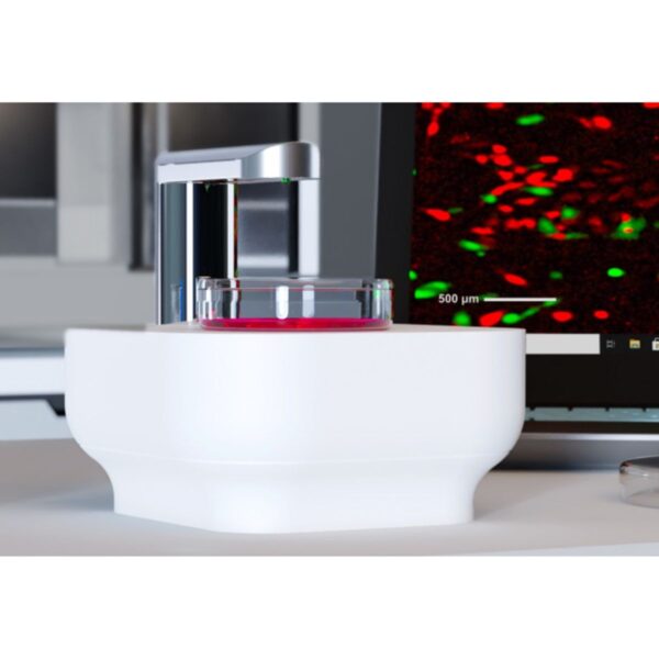
The Axion Lux3 FL is a small fluorescence live-cell imaging microscope equipped with one brightfield and two fluorescent channels (green and red). The device enables researchers to unravel cellular processes in real-time, provid…
The Axion Lux3 FL is a small fluorescence live-cell imaging microscope equipped with one brightfield and two fluorescent channels (green and red). The device enables researchers to unravel cellular processes in real-time, providing lots of valuable data about the experiments and answers if, when, how, and to what extend the cellular processes occur. Axion’s first fluorescent cell imaging device allows users to track dynamic cellular processes with high specificity by taking high-quality images to create real-time time-lapse movies. Simultaneously, the cells are kept in a controlled environment inside a standard cell culture incubator.
Let Lux3 FL capture fluorescent and brightfield images for you
Currently, fluorescent labeling is mostly used as an end-point measurement. However, time-lapse imaging of live cells can give much more information about biological processes. By using automated imaging at regular time intervals, the temporal resolution of the fluorescent data is increased, leading to even more relevant data about the cellular processes. In this way, with fluorescence microscopy, researchers can not only determine if a certain process has occurred, but also when it occurred and at what speed.