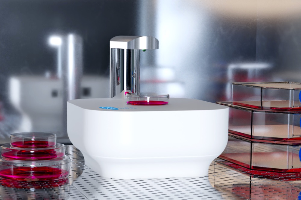
Axion – Automated Fluorescence Microscopy – Lux3 FL
The Axion Lux3 FL is a small fluorescence live-cell imaging microscope equipped with one brightfield and two fluorescent channels (green and red). The device enables researchers to unravel cellular processes in real-time, providing lots of valuable data about the experiments and answers if, when, how, and to what extend the cellular processes occur. Axion’s first fluorescent cell imaging device allows users to track dynamic cellular processes with high specificity by taking high-quality images to create real-time time-lapse movies. Simultaneously, the cells are kept in a controlled environment inside a standard cell culture incubator.
Let Lux3 FL capture fluorescent and brightfield images for you
Currently, fluorescent labeling is mostly used as an end-point measurement. However, time-lapse imaging of live cells can give much more information about biological processes. By using automated imaging at regular time intervals, the temporal resolution of the fluorescent data is increased, leading to even more relevant data about the cellular processes. In this way, with fluorescence microscopy, researchers can not only determine if a certain process has occurred, but also when it occurred and at what speed.
Technical Specifications
| Channels | Brightfield, green and red fluorescent channels |
| Magnification | 10× fixed objective – 20× digital zoom |
|---|---|
| Fluorescence filters | Green – excitation: 452/45 nm; emission: 512/23 nm Red – excitation: 561/14 nm; emission: 630/90 nm |
| Camera | 6.4 MP CMOS |
| Light source | LED |
| Data formats | JPG, TIFF, XLSX, MP4 |
| Image size | 2072 × 2072 pixels |
| Field of view | 1.45 mm × 1.45 mm |
| Culture vessels | Well-plates, petri dishes, flasks, microfluidic chips, and custom culture vessels |
| Computer requirements | Windows 10, 2.4 GHz i5 processor, 4 GB RAM memory, USB 3.0, internet |
| Dimensions (L × W × H) | 166 mm × 140 mm × 135 mm |
| Weight | 1.3 kg |
| Operating conditions | 5 – 40 °C, 20 – 95% humidity |
| Warranty | 1-year |
| Data storage | Unlimited cloud storage |
| Extra features | Template functionality (ability to save image acquisition parameters in templates), Object count (count the number of fluorescent objects in the image) |
Key Features

Live insight in cellular processes
Get the whole story and observe the development of cellular processes while they occur. The Axion Lux3 fluorescence imaging microscope automatically creates time-lapse movies that contain many cellular features. The videos are made from inside the incubator without disturbing your cells and can be immediately accessed and analyzed remotely via the Axion Cloud, providing real-time updates on your cell cultures and running experiments.

NEW – Quantification of real-time cellular events
For certain experiments, in addition to monitoring cell cultures, it is also required to quantitatively evaluate the progression of cellular events. The Axion Lux3 FL, equipped with a new Object Count algorithm, can automatically calculate the number of fluorescent objects in the image and investigate how this number changes over time. What’s more, the existing software interface has become even more intuitive, allowing users to quickly and easily adjust the parameters of an image.
Analyze cells in their desired culture environment
Kinetic fluorescence imaging of cellular processes requires environmental control throughout the experiment. Because of its small size, the Lux3 FL fits inside any standard incubator. This enables you to perform fluorescent live-cell imaging experiments parallel to your regular cell cultures. Because of the remote data accessibility in the Axion Cloud, your cells do not have to be disturbed during the experiment.
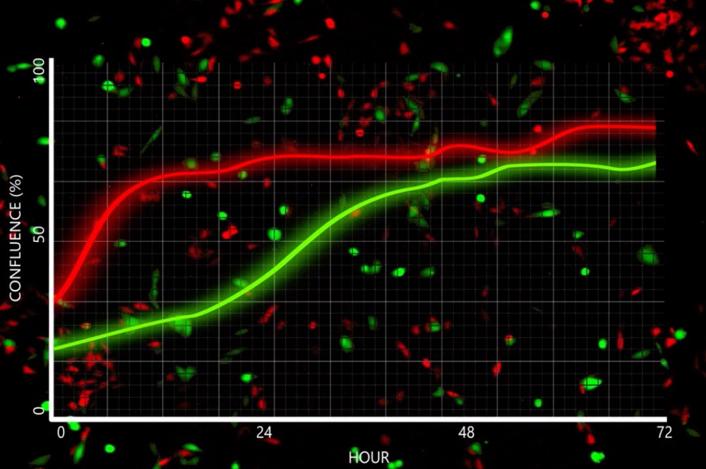
Simultaneous analysis of multiple cell features
Using Axion Lux3 FL, you can analyze green and red fluorescent tags simultaneously with your brightfield images. This not only saves you time but also provides tools to distinguish cells from each other or distinguish labeled structures within the cells. By tracking changes in the fluorescent signal over time, cell number changes or fluorescently tagged cellular components can easily be identified.
How To Use the Axion Lux3 FL Fluorescence Live-Cell Imager
Axion Lux3 FL Duo Kit – Side-By-Side Fluorescence Live-Cell Imaging
Axion Multi Lux3 FL – Simultaneous Fluorescence Live-Cell Imaging
อุตสาหกรรมหลัก
- ชีวเภสัชกรรมและเทคโนโลยีชีวภาพ